Sonohysterography (सोनोहिस्टेरोग्राफी), also called Hysterosonography, uses sound waves to produce pictures of the inside of the uterus and help diagnose many problems, including unexplained vaginal bleeding, infertility, and repeated miscarriages. Hysterosonography is performed very much like a gynecologic exam. Your doctor will insert a speculum into your vagina and insert a catheter into the cavity of the uterus. Using a small tube inserted into the vagina, your doctor will inject a small amount of sterile saline into the cavity of the uterus and study the lining of the uterus using the ultrasound transducer. Ultrasound does not use ionizing radiation, has no known harmful effects, and provides a clear picture of soft tissues that don’t appear well on X-rays.
What is Sonohysterography?
The word Sonohysterography can be broken down into parts to understand its meaning: “Sono-” = sound (refers to ultrasound, which uses sound waves to create images). “Hystero-” = uterus (from Greek hystera). “-graphy” = process of recording or imaging. So, Sonohysterography means: “Imaging of the uterus using sound waves.”

What are some common uses of Sonohysterography?
Sonohysterography is commonly used to evaluate the uterine cavity and is especially helpful when standard ultrasound doesn’t provide enough detail. Here are some of the most common uses of the procedure:
Investigating Abnormal Uterine Bleeding
-
Irregular periods
-
Heavy menstrual bleeding
-
Bleeding after menopause
-
Sonohysterography can help identify structural causes like polyps, fibroids, or a thickened endometrium.
Evaluating Infertility or Recurrent Miscarriages
-
It checks for uterine abnormalities (e.g., septate uterus, adhesions, or scarring) that might interfere with conception or carry a pregnancy to term.
Detecting Uterine Polyps or Fibroids
-
These growths may not be visible on a regular ultrasound.
-
Saline infusion opens the uterine walls to highlight any masses inside the cavity.
Assessing the Endometrium
-
This is especially important in postmenopausal women or those on hormone therapy.
-
Helps determine if the endometrial lining is too thick or irregular.
Guiding Further Procedures
-
It can guide decisions about whether a hysteroscopy or surgical treatment is needed.
-
Sometimes used before in vitro fertilization (IVF) to ensure the uterus is healthy for embryo transfer.
How should I prepare for Sonohysterography?
Preparing for Sonohysterography is usually pretty simple, but there are a few important things to keep in mind to ensure the procedure goes smoothly and comfortably:
When to Schedule It
-
The test is usually done after your period ends but before ovulation (typically days 5 to 10 of your cycle).
-
This timing avoids interfering with a possible pregnancy.
-
It also ensures the endometrium is thin for better visualization.
-
How to Prepare
-
Inform your doctor if:
-
You might be pregnant
-
You have a pelvic infection or a history of one
-
You have allergies (especially to latex or iodine, if used)
-
-
Medications:
-
You may be advised to take an over-the-counter pain reliever (like ibuprofen) 30–60 minutes before the procedure to reduce cramping.
-
-
Empty your bladder just before the test.
-
A transvaginal ultrasound requires a mostly empty bladder for better imaging.
-
-
No special fasting or diet is required.
Hygiene and Personal Care
-
You can shower or bathe as normal.
-
Avoid using tampons, douches, or vaginal medications the day of the exam unless directed otherwise.
What to Wear
-
Wear something comfortable and easy to remove, as you’ll change into a gown for the procedure.
What does the Sonohysterography equipment look like?
Great question! The Sonohysterography setup is pretty simple and uses equipment you’re likely already familiar with from regular pelvic exams and ultrasounds. Here’s what the main components look like:
Transvaginal Ultrasound Probe
-
A thin, wand-like device (about the size and shape of a tampon)
-
Covered with a protective sheath (like a condom) and lubricated
-
This is inserted into the vagina to create clear images of the uterus
Saline Infusion Catheter
-
A very thin, flexible tube (like a soft straw)
-
Inserted through the cervix into the uterus
-
Connected to a syringe or saline bag that delivers sterile saltwater into the uterine cavity
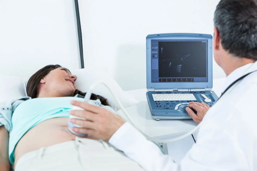
Ultrasound Machine
-
A standard ultrasound monitor that displays live images
-
The technician or doctor uses it to watch the uterus in real time as saline fills it and expands the cavity
Exam Table with Stirrups
-
Similar to what you’d see in a gynecologist’s office
-
You’ll lie back with your feet in stirrups during the procedure
Extras
-
Sterile gloves, lubricant, antiseptic wipes, and other disposable supplies for hygiene and comfort

How does the Sonohysterography procedure work?
Here’s a step-by-step breakdown of how the Sonohysterography procedure works, so you know exactly what to expect:
Preparation
-
You’ll be asked to undress from the waist down and lie on an exam table with your feet in stirrups (just like during a Pap smear).
-
A speculum is inserted into the vagina to open it gently.
Cleaning and Catheter Insertion
-
The cervix is cleaned with an antiseptic solution.
-
A thin, soft catheter (saline tube) is inserted through the cervix into the uterine cavity.
-
The speculum is then removed.
Transvaginal Ultrasound Probe
-
The doctor inserts a transvaginal ultrasound probe into the vagina.
-
The probe gives real-time images of your uterus on a monitor.
Saline Infusion
-
Sterile saline is slowly infused through the catheter into the uterus.
-
The saline expands the uterine cavity, separating the walls and making it easier to spot any abnormalities like polyps, fibroids, or scarring.
Imaging and Evaluation
-
As the saline fills the uterus, the ultrasound captures detailed images of:
-
The uterine lining (endometrium)
-
The shape and contour of the uterine cavity
-
Any abnormal growths or structural issues
-
Completion
-
Once the imaging is done, the saline and catheter are removed.
-
The entire procedure usually takes 10 to 20 minutes.
After the Procedure
-
You may have mild cramping, watery discharge, or spotting for a day.
-
Most people return to normal activities immediately.
How is the Sonohysterography procedure performed?
Here’s a detailed explanation of how the Sonohysterography procedure is performed, step by step:
Patient Positioning
-
Like a pelvic exam, you’ll lie on an exam table with your feet in stirrups.
-
A speculum is inserted into the vagina to allow access to the cervix.
Cervix Cleaning and Catheter Insertion
-
The cervix is gently cleaned with an antiseptic solution.
-
A thin, soft catheter (usually around 1–2 mm wide) is inserted through the cervix into the uterus.
-
The speculum is then removed, and the catheter remains in place.
Transvaginal Ultrasound Begins
-
A transvaginal ultrasound probe (a slim wand-shaped device) is inserted into the vagina.
-
It provides clear, real-time images of the uterus on a monitor.
Saline Infusion
-
Sterile saline (salt water) is slowly injected through the catheter into the uterus.
-
The saline expands the uterine cavity, helping separate the walls and highlighting any internal abnormalities (e.g., polyps, fibroids, adhesions).
Image Capture and Evaluation
-
While the saline is present in the uterus, the ultrasound captures detailed images of the:
-
Endometrial lining
-
The shape of the cavity
-
Any abnormal structures inside
-
-
The doctor evaluates the findings in real-time and may save key images for your records.
Completion
-
The saline, catheter, and ultrasound probe are all gently removed.
-
The entire procedure usually lasts 10 to 20 minutes.
What to Expect Afterward
-
Mild cramping or light spotting for a few hours is common.
-
You can return to normal activities on the same day.
What are the experiences during and after the procedure?
Here’s what most people typically experience during and after a Sonohysterography—so you can feel more prepared and less anxious:
During the Procedure
Common Experiences
-
Mild to moderate cramping (like menstrual cramps), especially when:
-
The catheter is inserted
-
The saline is infused into the uterus
-
-
Pressure or fullness in the lower abdomen
-
Some discomfort from the transvaginal ultrasound probe or speculum
What Helps
-
Taking an over-the-counter pain reliever (like ibuprofen) before the appointment can ease cramping.
-
Deep breathing or relaxing your pelvic muscles during the exam can reduce discomfort.
Most people say the procedure is uncomfortable, but tolerable, and it usually lasts about 10–20 minutes.
After the Procedure
Common Post-Procedure Effects
-
Mild cramping for a few hours
-
Watery discharge (from the saline) that may be tinged with a little blood
-
Light spotting for up to a day
Wearing a panty liner after the procedure is a good idea.
When to Call Your Doctor
-
Heavy bleeding
-
Fever or chills
-
Severe or increasing pain
-
Foul-smelling discharge (could signal infection)
Recovery
-
You can resume normal activities immediately unless your doctor tells you otherwise.
-
Most people return to work or their regular routines the same day.
What are the benefits of Sonohysterography?
Sonohysterography offers several key benefits, especially when it comes to diagnosing uterine issues with precision and minimal discomfort. Here’s a breakdown of the advantages:
Main Benefits of Sonohysterography
Clearer Visualization of the Uterus
-
The saline expands the uterine cavity, separating the walls and allowing better imaging.
-
Helps detect abnormalities like polyps, fibroids, adhesions, or congenital defects that might not show up clearly on standard ultrasound.
Noninvasive and Low Risk
-
Unlike a hysteroscopy (which involves a camera), this is minimally invasive.
-
No incisions, no anesthesia, and very low risk of complications.
More Accurate than Standard Ultrasound
-
Offers superior detail of the endometrial lining and cavity.
-
Can reveal structural causes of abnormal bleeding or infertility that a regular transvaginal ultrasound might miss.
Helpful in Infertility Workups
-
Detects uterine issues that could interfere with implantation or pregnancy.
-
Useful before procedures like IVF, to ensure the uterus is healthy and receptive.
Quick and Convenient
-
The procedure usually takes 10–20 minutes.
-
Done in an outpatient setting, with minimal recovery time.
Guides Further Treatment
-
Helps doctors decide whether more invasive steps like hysteroscopy or surgery are needed.
-
It can reduce unnecessary procedures by clarifying the diagnosis early.
What are the drawbacks of Sonohysterography?
Sonohysterography (SHG), also known as saline infusion sonography (SIS), is a diagnostic imaging procedure used to examine the uterus, particularly the endometrial cavity, for abnormalities. While it’s a useful and minimally invasive tool, there are some potential drawbacks:
Discomfort or Pain
Some women experience discomfort or mild pain during the procedure, as the saline is introduced into the uterus. The pressure from the saline infusion can also cause cramping.
Risk of Infection
Though rare, there is a slight risk of infection when introducing saline into the uterus. This can lead to pelvic inflammatory disease (PID) or other infections if not done under sterile conditions.
Allergic Reactions
There is a very small risk of an allergic reaction to the contrast material used during the procedure. Though saline is generally considered safe, any foreign substance can occasionally cause a reaction.
Inability to Detect Certain Conditions
While SHG is effective at detecting polyps, fibroids, and abnormalities in the endometrium, it may not detect conditions affecting deeper layers of the uterus or certain types of cancer.
Limited to Certain Cases
Sonohysterography is primarily used for women with abnormal bleeding or those undergoing infertility evaluations. It is not suitable for all gynecological conditions, such as those requiring a detailed look at the ovaries or external pelvic structures.
Contraindications
SHG is not recommended for women with active pelvic infections, those who are pregnant, or those with certain uterine abnormalities (e.g., large fibroids) that may affect the quality of the imaging.
False Positives or False Negatives
As with any diagnostic test, there’s a chance of false positives or false negatives, where the test either identifies a problem that isn’t there or misses an actual issue.
Overall, while sonohysterography is a relatively safe and non-invasive procedure, these factors should be taken into account when considering it as a diagnostic tool.
Conclusion
In conclusion, sonohysterography (SHG) is a valuable diagnostic tool in gynecology, offering a non-invasive and effective way to evaluate the uterine cavity for abnormalities such as polyps, fibroids, and other structural issues. It is particularly useful for women experiencing abnormal uterine bleeding or those undergoing fertility evaluations. While the procedure is generally safe and well-tolerated, it is not without drawbacks, including potential discomfort, the risk of infection, and limitations in detecting certain conditions. Additionally, SHG is not suitable for all patients, and its results can sometimes lead to false positives or negatives. Despite these limitations, sonohysterography remains a crucial part of modern gynecological care, providing valuable insights into uterine health when used appropriately.
Frequently Asked Questions
1. What is sonohysterography (SHG)?
Sonohysterography (SHG), also known as saline infusion sonography (SIS), is a type of ultrasound procedure used to examine the inside of the uterus. A sterile saline solution is infused into the uterine cavity, and ultrasound imaging is used to detect abnormalities such as polyps, fibroids, or uterine anomalies.
2. Why is sonohysterography performed?
SHG is typically performed to investigate causes of abnormal uterine bleeding, infertility, recurrent miscarriages, or to evaluate the uterine cavity for structural issues like polyps, fibroids, or adhesions that might affect fertility or overall uterine health.
3. How is the procedure performed?
The procedure involves inserting a thin catheter through the cervix into the uterus, where a sterile saline solution is introduced. The doctor uses ultrasound to visualize the uterus and assess the shape and structure of the uterine cavity.
4. Is sonohysterography painful?
Most women experience mild discomfort or cramping during the procedure as the saline is introduced into the uterus. The discomfort usually subsides once the procedure is completed, but the level of pain varies from person to person.
5. Is sonohysterography safe?
Sonohysterography is generally considered safe. The procedure uses ultrasound and saline, both of which are non-invasive and low-risk. However, there is a small risk of infection, allergic reaction to the saline, or minor bleeding after the procedure.
6. Are there any risks associated with sonohysterography?
While rare, potential risks include infection, allergic reactions to the saline, and uterine injury. It is also not recommended for women with active pelvic infections or those who are pregnant.
7. How long does the sonohysterography procedure take?
The procedure typically takes about 15 to 30 minutes. The actual insertion of the catheter and saline infusion usually takes just a few minutes, while the rest of the time is spent capturing and reviewing the ultrasound images.
8. What should I expect after the procedure?
After SHG, you may experience mild cramping or spotting for a few hours to a day. It’s advised to avoid sexual intercourse, tampon use, or douching for a short period after the procedure to reduce the risk of infection.
9. Can sonohysterography detect all uterine conditions?
While SHG is effective in detecting abnormalities within the uterine cavity, such as fibroids, polyps, and adhesions, it may not be able to identify conditions that affect the deeper layers of the uterus or outside structures. It also does not provide detailed information about the ovaries.
10. How soon will I get the results of sonohysterography?
The results of the procedure are usually available within a few days. The images taken during the SHG are reviewed by a radiologist or the attending physician, and a follow-up appointment will be scheduled to discuss the findings.
Reference: https://www.radiologyinfo.org/en/info/hysterosono